Cat. No. | Description | Pcs./Box |
|
|---|
81126 | µ-Slide I 0.1 Luer, ibiTreat, tissue culture treated, sterile | 15 |
| 81122 | µ-Slide I 0.1 Luer, Collagen IV, sterile | 15 |
| 81123 | µ-Slide I 0.1 Luer, Fibronectin, sterile (available only on request) | 15 |
| 81121 | µ-Slide I 0.1 Luer, hydrophobic, uncoated, sterile | 15 |
| 80166 | µ-Slide I 0.2 Luer, ibiTreat, tissue culture treated, sterile | 15 |
| 80162 | µ-Slide I 0.2 Luer, Collagen IV, sterile | 15 |
| 80163 | µ-Slide I 0.2 Luer, Fibronectin, sterile (available only on request) | 15 |
| 80164 | µ-Slide I 0.2 Luer, Poly-L-Lysine, sterile | 15 |
| 80165 | µ-Slide I 0.2 Luer, Poly-D-Lysine, sterile (available only on request) | 15 |
| 80161 | µ-Slide I 0.2 Luer, hydrophobic, uncoated, sterile | 15 |
| 80176 | µ-Slide I 0.4 Luer, ibiTreat, tissue culture treated, sterile | 15 |
| 80172 | µ-Slide I 0.4 Luer, Collagen IV, sterile | 15 |
| 80173 | µ-Slide I 0.4 Luer, Fibronectin, sterile (availabel only on request) | 15 |
| 80174 | µ-Slide I 0.4 Luer, Poly-L-Lysine, sterile | 15 |
| 80175 | µ-Slide I 0.4 Luer, Poly-D-Lysine, sterile (available only on request) | 15 |
| 80171 | µ-Slide I 0.4 Luer, hydrophobic, uncoated, sterile | 15 |
| 80186 | µ-Slide I 0.6 Luer, ibiTreat, tissue culture treated, sterile | 15 |
| 80182 | µ-Slide I 0.6 Luer, Collagen IV, sterile | 15 |
| 80183 | µ-Slide I 0.6 Luer, Fibronectin, sterile (available only on request) | 15 |
| 80184 | µ-Slide I 0.6 Luer, Poly-L-Lysine, sterile | 15 |
| 80185 | µ-Slide I 0.6 Luer, Poly-D-Lysine, sterile (available only on request) | 15 |
| 80181 | µ-Slide I 0.6 Luer, hydrophobic, uncoated, sterile |
|
| 80196 | µ-Slide I 0.8 Luer, ibiTreat, tissue culture treated, sterile | 15 |
| 80192 | µ-Slide I 0.8 Luer, Collagen IV, sterile | 15 |
| 80193 | µ-Slide I 0.8 Luer, Fibronectin, sterile (available only on request) |
|
| 80194 | µ-Slide I 0.8 Luer, Poly-L-Lysine, sterile |
|
| 80195 | µ-Slide I 0.8 Luer, Poly-D-Lysine, sterile (available only on request) |
|
| 80191 | µ-Slide I 0.8 Luer, hydrophobic, uncoated, sterile |
|
|
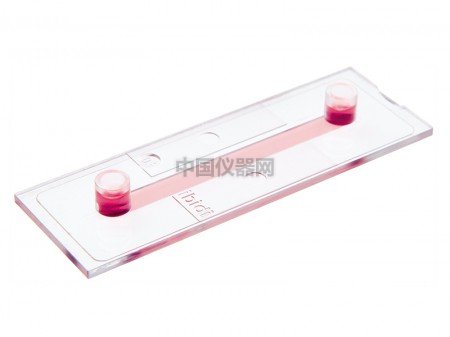
Channel slides with different heights, volumes, and coatings specially suited for flow applications---ibidi
o Large area of uniform shear stress
o Easy connection using Luer adapters
o Homogeneous cell distribution with defined optical pathway
Applications:
o Adherent cells under flow conditions
o Cell culture (static or stop-flow)
o 3D cell culture in gels brought into the channels
o High-resolution microscopy of living and fixed cells
Specifications:
Channel length |
50 mm |
Adapters |
Female Luer |
Volume per reservoir |
60 μl |
Growth area per channel |
2.5 cm2 |
Bottom: ibidi Standard Bottom |
Technical Features: o Standard format with thin bottom for low or high magnification microscopy (up to 100x) o Large observation area for microscopy o Channel volumes of 25 µl, 50 μl, 100 μl, 150 μl, or 200 μl o Defined shear stress and shear rate levels o Easy connection to tubes and pumps using Luer adapters o Available as variety pack containing all heights o Also available as a Flow Kit with tubing and adapters o Fully compatible with the ibidi Pump System
Cross Section of the Channel: Same Slide – Different Channel Height and VolumeCross Section of the Channel: Same Slide – Different Channel Height and Volume Choosing the Right Channel for Flow ApplicationsFor flow assays with small amounts of medium and high values of shear stress | 0.1 and 0.2 mm
channels | | For a wide range of shear stress | 0.4 mm channel | | For controlling low values of shear stress (<2 dyne /cm²) | 0.6 and 0.8 mm
channels |
Static Cultures vs. Flow ApplicationsThe general rule is:
Low channels are more suitable for flow applications.
High channels are more suitable for static cell culture
Compatible with Solvents for Staining and Fixation HUVEC, cultivated over 7 days at 10 dyn / cm². VE-cadherins are stained in green, cell nuclei are stained in blue.
Influence of Shear Stress on Cultured Cells Human umbilical vein endothelial cells (HUVEC) cultured under flow conditions (20 dyn / cm²) in a µ-Slide I 0.4 Luer over 9 days. The primary cells were transduced with the adenoviral vector rAV CMV-LifeAct-TagRFP 24 hours prior to the experiment. |